The power and flexibility to create any event registration you can imagine

Create any event, simple or complex
Customise not only the look and feel, but the flow of your registration. Add configurable registration paths for more personalisation.

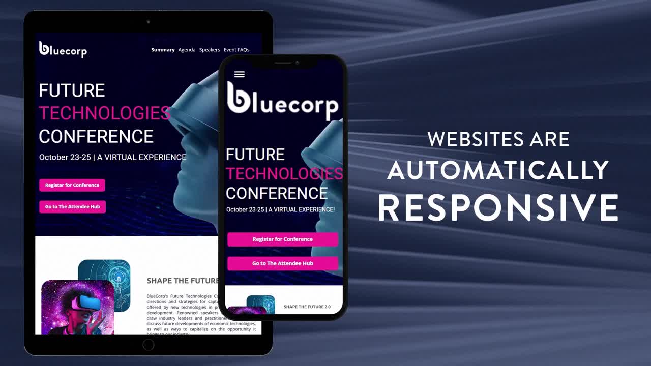
Build a website that wows your audience
Create fully branded event experiences with drag-and-drop widgets, or take it to the next level with custom CSS classes.

Increase conversions and drive registrations
Capture attendees with high intent, monitor abandoned registrations, send reminders, and create tasks for sales.
Frequently Asked Questions
Signing up is easy. Simply click HERE and we’ll immediately send you an email so you can activate your account.
Anyone can sign up for a trial. If you are already a Cvent Event Management customer, check your account first as you may already have access to the functionality available in the trial.
Access to, and usage of, Cvent services are governed by Cvent’s Terms of Use, available HERE
There are no geographic restrictions for trial sign up. The trial supports multiple languages and currencies.
No. It’s free to sign up for the trial and to build your events. Launching your event is also free as you will only be charged when you capture registrations.
We ask customers to provide their credit card information as a backup in the event that what they’re charging does not cover our fees (registration fee & revenue share).
Credit cards will be charged only when you have a negative balance. For example, if the cost of registration doesn’t cover our fees, such as in the case of a free event, the credit card will be charged.
After you build your event and click the “Launch Event” button, you will be presented with your payment options. Our pay-as-you-go payment option consists of a per registration fee, a small percentage of revenue share, and a Cvent Payment Services fee should you decide to use our payment services.
Alternatively, after you’ve completed the Event Build Wizard and exited the initial Site Designer, you’ll be able to see your account information. If you go to Admin à Account à Overview, you’ll see the pricing information listed there.
To use the Cvent Professional Licence with the pay-as-you-go payment option, all you need to do is click “Launch Event”, enter your billing information and your event will become live instantly. You will only pay when people sign up for your event.
If you’d like more information about our contract options, call us at 800-925-7220 to speak with a Cvent sales professional.
Void where prohibited by law. Subject to cancellation or change at any time without prior notice.
Additional terms may apply. All rights reserved.